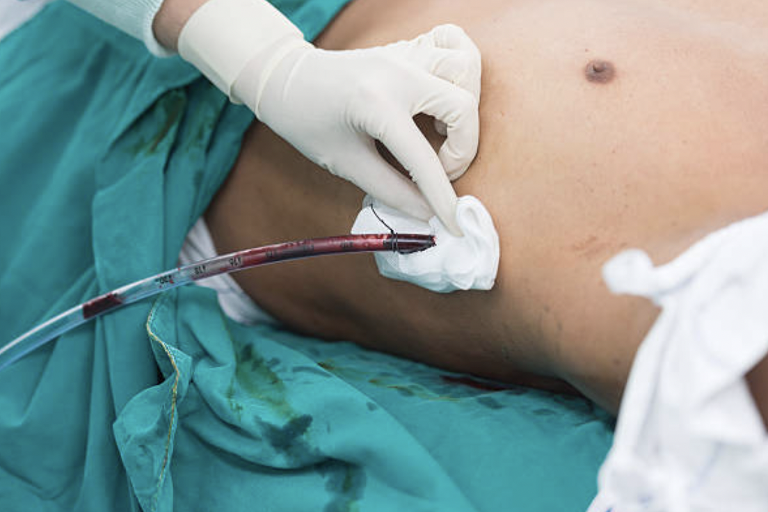
Chest Drain

Here's a quick run-through of the key points to know for this procedure, which can crop up at either level in the FRCA examination.
Indications
- Pneumothorax*
- Pleural effusion
- Haemothorax
- Empyema
- Chylothorax
*Either traumatic, post-operative, as a result of invasive ventilation, or a tension pneumothorax that has already been decompressed with a needle.
Contraindications
- Nil absolute
Relative
- Coagulopathy
- Local infection
Complications
- Failure
- Pain
- Haemorrhage
- Trauma to surrounding structures
- Pneumothorax
- Misplaced drain (including extra-pleural placement which may appear correct on a chest xray)
- Infection
- Blocked drain
- Surgical emphysema
Equipment
- Sterile drape and chest drain kit
- Lidocaine
- Chest drain
- Scalpel
- Artery forceps
- Blunt forceps
- Sutures
- Needle holder
- Gauze and dressing
- Underwater seal
- Sterile water
Site
Anterior approach
- Second intercostal space
- Mid clavicular line
- Lateral to internal mammary artery
- Better for apical air drainage
Lateral approach
- Fifth or sixth intercostal space
- Mid axillary line
- Better for fluid
- More likely to hit liver or diaphragm
Procedure
- Monitoring
- Confirm consent, identity, indications and contraindications
- Adequate skilled assistance
- Confirm diagnosis and site with ultrasound where possible
- Sterile technique
- Local anaesthetic
- Skin incision of 2-3cm parallel to ribs in intercostal space, just superior to rib edge
- Introduce closed forceps through incision into the pleural space
- Sweep with finger to confirm pleural cavity entered and to check for lung tissue and adhesions
- Insert drain using forceps
- Aim cranially if draining air, posteriorly if draining fluid
- Connect to underwater seal and confirm 'swing' in drain
- Purse suture to secure drain
- Dressing
- Xray to confirm
Afterwards
- Ensure drain kept below level of patient
- Serial xrays to monitor progress and confirm placement
Indications to remove drain
- No longer draining air or fluid
- Lungs fully expanded on xray
- No significant leak on coughing or valsalva manoeuvre
- Some say to clamp drain for 24h then re-xray to confirm no reaccumulation
Contraindications
- IPPV
Complications
- Reaccumulation
- Tension pneumothorax
Here's a really useful video
Cool tweets
PSA: this is why we ALWAS scan with colour doppler (or CPD, in this case) to look for vessels prior to thoracentesis.
— Katie Wiskar (@katiewiskar) March 29, 2023
Indicator is toward patient's head, so the vessel is running on the superior aspect of the rib --> right where you'd normally poke #POCUS #MedEd pic.twitter.com/7SwCl6FgUM
Pulmonary teaching case: you are called to the bedside of a 60yo man who was admitted for pneumonia a week ago. You were called because “he coughed and now his chest is PULSATING!”
— Nick Mark MD (@nickmmark) May 28, 2023
This is what you see at the site of a previously removed chest drain:
What’s the diagnosis?
1/ pic.twitter.com/BlBOFcyIL9
🧵regarding some technical points about passing drains or chest tubes through the body wall.
— Ron Barbosa MD FACS (@rbarbosa91) May 19, 2023
Aside from the normal techniques, we will show what to do with the less common 'trocar' type of drains and chest tubes, and also what the 'pointy' part of the chest tube is for.
(1/ ) pic.twitter.com/LhbWd3STd7
